Cell-free expression system offers advantages over cell-based expression systems for difficult-to-express proteins, not only for membrane proteins, but also for proteins with multiple disulfide bonds or cytotoxic proteins, for example.
Firstly, proteins that are toxic for the production host are often difficult to express in high yields. Cell-free expression system overcomes this issue because cell growth and viability are not required for protein expression. Therefore, the cell-free system enables, for example the production of cytotoxic cancer therapeutic proteins, such as Pseudomonas exotoxin (PE)-based immunotoxins, targeting tumor-related antigens expressed in various cancers (Weldon et al., 2011; Wolf et al., 2009). Antimicrobial peptides and proteins can also be expressed in cell-free systems where they induce bacterial death or growth inhibition in cell-based systems (Jin et al., 2018).
Additionally, the production of functional plasma membrane proteins can be enhanced due to the open nature of the cell-free system that allows a unique reaction environment control by supplementing key components at precise concentrations. Such reaction is not possible in cell-based systems. (Jin et al., 2018). Indeed, the formation of disulfide bonds is promoted by optimizing the redox potential and by adding chaperones and isomerases (Carlson et al., 2012; Rosenblum et al., 2014). Protein aggregation can also be prevented by the addition of stabilizing agents, such as polyethylene glycols, polysaccharides and amino acids (Kai et al., 2013).
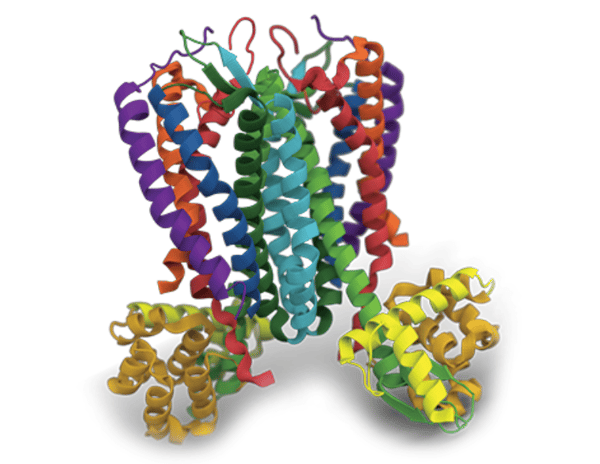
Finally, cell-free expression systems have been largely exploited to produce plasma membrane proteins that are an important class of drug targets. Due to their complex structures and hydrophobic transmembrane domains, cell membrane proteins are difficult-to-express in cell-based systems. In contrast, cell-free open systems offer the flexibility to supplement reactions with detergents or lipids that mimic membrane structures and therefore help solubilizing membrane proteins (Carlson et al., 2012; Jin et al., 2018). Since membrane proteins behave differently, the systematic screening of the expression environment is important, starting with detergent screening. However, numerous membrane proteins do not fold correctly in presence of detergent and require lipids to support their functional folding and stability. Thus, well-defined and preformed bilayers, such as liposomes and nanodiscs can be added into the cell extract to be present when these types of protein are being expressed (Henrich et al., 2015).
With such approach, Synthelis has produced more than 150 cell membrane proteins including about 20 GPCRs in a proteoliposome format, using its patented cell-free expression system. Most of the expressed GPCRs have shown a proper folding assessed either by Surface Plasmon Resonance (SPR) analysis, by ELISA or by radio-ligand binding assay.
As an example the CXC Chemokine Receptor 4 (CXCR4) was produced at Synthelis at up to 10 mg quantities in a proteoliposome format. The proper folding of CXCR4 protein was validated by SPRi (SPRiPlex, Horiba) and BLI (Octet RED96, Pall – Fortebio) with 4 different ligands (CXCL12, T22, AMD 3100 and 12G5 antibody). Immunization campaigns were then successfully performed with those functional CXCR4-containing proteoliposomes in lamas as well as in mice. As a result, specific antibodies against CXCR4 expressing HEK cells were obtained by Fair Journey Biologics, when the PRC Team of the INRA center of Nouzilly selected a strong CXCR4 antagonist when using CXCR4 proteoliposomes in their immunization and their phage display process. In the same way, nanodiscs have been demonstrated to be efficient for the insertion of G-protein-coupled receptors (GPCRs) (Gao et al., 2012; Proverbio et al., 2013) when incorporated into cell-free systems
As shown in this article and supported by a growing list of scientific publications, the power of cell-free systems lies in their ability to bypass the biological limitations of the cells and express difficult proteins, not only integral membrane proteins, but also cytotoxic, complex, instable or insoluble ones.
Authors & sources :
Carlson E.D., Gan R., Hodgman C.E., Jewett M.C. 2012. Cell-free protein synthesis: Applications come of age. Biotechnology Advances 30:1185–1194.
Henrich E., Hein C., Dötsch V., Bernhard F. 2015. Membrane protein production in Escherichia coli cell-free lysates. FEBS Letters 589:1713–1722.
Gao T., Petrlova J., He W., Huser T., Kudlick W., Voss J., Coleman M.A. 2012. Characterization of de novo synthesized GPCRs supported in nanolipoprotein discs. PLoS One 7(9), e44911.
Isaksson L., Enberg J., Neutze R., Karlsson B.G., Pedersen A. 2012. Expression screening of membrane proteins with cell-free protein synthesis. Protein Expression and Purification 82:218–225.
Jin X., Hong S.H. 2018. Cell-free protein synthesis for producing ‘difficult-to-express’ proteins. Biochemical Engineering Journal 138:156–164.
Kai, L., Dötsch, V., Kaldenhoff, R., Bernhard, F. 2013. Artificial environments for the co-translational stabilization of cell-free expressed proteins. PLoS ONE 8(2): e56637.
Proverbio D., Roos C., Beyermann M., Orbán E., Dötsch V., Bernhard F. 2013. Functional properties of cell-free expressed human endothelin A and endothelin B receptors in artificial membrane environments. Biochim. Biophys. Acta 1828(9):2182–2192.
Rosenblum G. and Cooperman, B.S. 2014. Engine out of the chassis: Cell-free protein synthesis and its uses. FEBS Letters 588:261–268.
Weldon J.E., Pastan I. 2011. A guide to taming a toxin: recombinant immunotoxins constructed from Pseudomonas exotoxin A for the treatment of cancer, FEBSJ. 278:4683–4700.
Wolf P., Elsässer-Beile U. 2009. Pseudomonas exotoxin A: from virulence factor to anti-cancer agent, Int. J. Med. Microbiol. 299:161–176.


